Saturday, June 9, 2012

Type II Diabetes: It’s Not Just a Human Problem!


Type II diabetes (T2D) is a metabolic disorder characterized by hyperglycemia (high blood glucose levels), insulin resistance, and a host of other symptoms such as obesity, poor circulation, and heart problems. It is the fifth leading cause of death in the United States, and the number of people diagnosed per year continues to increase.

Two of the defining characteristics of T2D, hyperglycemia and insulin resistance, are directly related to each other. Insulin is a hormone secreted by the pancreas that causes cells to take up glucose from the blood, where it is used for energy. In T2D patients, the body’s cells are desensitized to insulin; the cells do not respond adequately to normal levels of insulin, and as a result glucose remains the in the blood. This is the cause of hyperglycemia so closely associated with T2D!


Though studies suggest a strong link between high caloric diets with increasing rates of T2D (like eating too much sugar and simple carbohydrates!), human patients are rarely diagnosed with T2D because of their diet alone. In addition to diet, other complicating factors include lifestyle choices, stress, and genetic predisposition to the disease. Although the genetic factor of T2D is highly complex in humans, mutations in the genes controlling gluconeogenesis, β-oxidation, and insulin-dependant FOXO targets are consistently correlated with an increased chance of developing T2D.
  

A recent study published in 2011 suggests that fruit flies, Drosophila melanogastor , can also develop T2D with only a high sugar diet, and exhibit many of the same pathologies in metabolism that are seen in human patients with T2D! Like humans, Drosophila have an “insulin-like” hormone (DILP, Drosophila insulin-like peptide) that shares a very similar structure and function with human insulin. When fly larvae are raised on high sugar diets, they develop hyperglycemia and DILP resistance, despite being shown to have normal levels of DILP in their bodies.

The study also pinpointed potential causes of DILP-resistance in the flies, on a genetic level. To analyze for abnormal gene expression, RNA concentrations in high-sugar fed flies were compared with those in control flies. DNA is transcribed into RNA, which is then translated into the various proteins associated with the transcribed genes. Increased concentrations of one RNA transcript suggests elevated activity in its associated gene, while decreased levels suggests lower activity. The study found that the DILP-resistant cells had lower expression of an important T2D susceptibility gene hexokinase C, and increased levels of gluconeogenesis and B-oxidation; all of these observations are consistent with previous genetic studies in human patients with T2D!


A flowchart summarizing the hypothesized mechanism of insulin resistance in Drosophila.

Although the exact mechanism that causes insulin resistance is unclear, these metabolic processes have been well studied and are strongly associated with T2D in humans, mice, and now possibly Drosophila.  The implications of these results are quite revolutionary; they suggest that the metabolic mechanisms of T2D are evolutionarily conserved across vertebrates (humans) and invertebrates (Drosophila)! With the help of the fruit fly as an excellent and simple model for this common human disorder, we may be able to sort out the exact consequences of diet, lifestyle, and genetics separately in T2D development in humans. While human T2D is immensely complex, teasing out the nuances of each factor may hold the key to developing more effective treatments for patients in the future!


Connie Duong   


Tuesday, May 1, 2012

Are You What Your Mom Ate?


To ensure they give birth to a healthy baby, pregnant mothers will take the necessary precautions, such as eating a balanced diet or avoiding exposure to cigarette smoke. This is because there appears to be a link between exposure to environmental factors early in development and disease in adulthood. One explanation for this relationship may involve epigenetic modifications, such as DNA methylation of transposable elements.
The study featured in the article, Maternal nutrient supplementation counteracts bisphenol A-induced DNA hypomethylation in early development, sought to explore the effects of chemical exposure, specifically bisphenol A, during early development on the epigenome and whether or not these effects could be counteracted by nutrient supplementation.
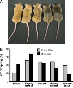
Epigenetic modifications established during early development create epialleles, or alleles that would be identical if not for methylation differences. One example of an epiallele is the Agouti gene in the yellow agouti mouse. The Avy allele of this gene resulted from the insertion of a retrotransposon into the 5' end of the gene. Methylation of cytosines in cytosine-guanine (CpG) dinucleotide sites in and near the Avy gene results in a wide variation in coat color, ranging from yellow (unmethylated) to pseudoagouti (methylated).
Bisphenol A (BPA) is a chemical used in the manufacture of polycarbonate plastics. It can be found in products such as food and beverage containers and baby bottles. Although reviews of the BPA literature have conflicting conclusions as to its potential as a human health risk, rodent studies have found that exposure to BPA, either as an embryo or fetus or as a newborn, is associated with higher body weight, increased breast and prostate cancer, and altered reproductive functions. The current study looked at the effect on the epigenome of offspring of maternal BPA exposure alone and in combination with nutritional supplements.
The study showed that maternal BPA exposure shifted the coat color distribution of genetically identical heterozygous offspring towards the yellow coat color phenotype. Twenty-one percent of offspring exposed to BPA during development were yellow compared with ten percent of offspring with no BPA exposure. Bisulfite treatment and sequencing was used to measure the DNA methylation at nine CpG sites in the promoter region of the Avy allele. The offspring that were exposed to BPA had 27+/- 2.8% methylation across the nine sites compared with 39 +/- 2.6% in the offspring without BPA exposure. The sites that exhibited the greatest difference in methylation appear to be important in modifying chromatin structure and Agouti gene expression. Early stem cell development must to be sensitive to BPA exposure since the patterning of DNA methylation at the Avy locus was similar in tissues from three of the germ layers (ectoderm, mesoderm, and endoderm).
Additionally, results from a statistical approach, known as mediational regression analysis, suggest that BPA exposure does not directly regulate coat color. Instead, coat color is determined by the methylation at the Avy gene. BPA exposure just so happens to affect methylation and therefore indirectly affects coat color.
The study investigated the relationship between BPA exposure and maternal nutritional supplements that are methyl donors, such as folic acid. Previous studies suggest that these supplements will counteract the hypomethylation effect of BPA exposure. Female mice that received a BPA diet supplemented with methyl donors or genistein (not a methyl-donating compound) produced offspring that exhibited the coat color distribution found in offspring from females that were not exposed to BPA. These findings suggest that maternal nutritional supplementation, with either methyl-donors or non-methyl-donors, counteracts the hypomethylation caused by maternal BPA exposure.
BPA exposure is linked to modifications of the epigenome and disease pathologies that are passed down through the germ line. This study’s findings suggest that nutrition interventions during early development have the potential to reduce disease susceptibility in adulthood. Be sure to thank your mom for eating healthy during her pregnancy!

Not So Identical


Diane Arbus, Identical Twins - Stencil                                       
Identical twins have largely the same genes. So, how can identical twins not look exactly the same or have the same susceptibility to disease? The phenotypic differences we see in twins prove that our genes are not the only factors in determining physical attributes. One explanation for the differences seen in identical twins is the presence of epigenetic differences.
The article, Epigenetic differences arise during the lifetime of monozygotic twinsfeatures a study which examined the difference in DNA methylation and histone acetylation between 80 identical twins over various ages. Identical twins are the perfect subjects to evaluate the epigenetic causes of phenotypes because the differences between the twin pairs must be a result of factors other than gene sequence.
The epigenome controls the differential expression of genes. DNA methylation and histone modifications, such as acetylation, store epigenetic information that controls heritable states of gene expression. In the article, the study examined different epigenetic characters, which included the abundance of DNA methylation and histone acetylation, as well as the distribution of DNA methylation in chromosomes.
Christoph Bock (Max Planck Institute for Informatics)
Histones are proteins that, together with DNA, are found in the chromatin in a cell's nucleus. This study examined the acetylation of two specific histones, H4 and H3,  as well as methylation of DNA. The technique known as Amplification of Intermethylated Sites (AIMS) was used to determine the specific methylated DNA sequences, which allowed for the determination of the distribution of DNA methylation on chromosomes.
35% of the twin pairs studied had significantly different epigenetic characters between pairs. The twins that were the most epigenetically different were older in age, while the youngest pairs were epigenetically similar. Also, twins with similar amounts of DNA methylation and histone acetylation, and methylation patterns, shared a common distribution of DNA methylation in their chromosomes. 
Twins by Elsie esq.

Using the statistical methods, ANOVA and ESD, the epigenetic variability was compared within the twin population and between pairs. Results from these methods determined that the epigenetic variability among individuals is high and similar, regardless of age group. However, older identical twins have higher epigenetic variability between pairs than younger twins. When comparing 3-year old twins with 50-year old twins, the 50-year-old twins were found to have extremely different gene expression. In an older twin pair, there were four times as many differently expressed genes than in the younger twin pair.
Besides age, other factors that were found to possibly contribute to the epigenetic modification pattern differences between twin pairs included the time twins spent with each other and differences in their medical history. Twins who spent the least amount of their lifetimes together or had different medical history were those who also showed the greatest differences in levels of DNA methylation and histone acetylation of histones. Even when comparing different cell types (epithelial mouth cells, intraabdominal fat, and skeletal muscle biopsies), there are striking epigenetic differences in older twins with different lifestyles and that had spent less of their lives together.
twins by cesarastudillo
The difference in epigenetic patterns that arise during the lifetime of identical twins explains their phenotypic differences. External factors, such as smoking habits, physical activity, or diet, have been proposed to have a long-term influence on epigenetic modifications. Internal factors, such as small defects in transmitting epigenetic information through successive cell divisions, may also play a role in causing different epigenetic modification patterns. Further studies must be done to fully understand the effects of these external and internal factors on the the epigenome. We have much more to learn about the growing research field of epigenetics and how different phenotypes can result from the same genotype.